what allows the ribs to attach to the thoracic vertebrae
| Rib cage | |
|---|---|
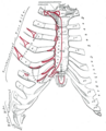
 The human rib cage. (Source: Gray'south Beefcake of the Human Trunk, 20th ed. 1918.) | |
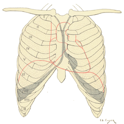 Protection on the rib muzzle of the heart, lungs and diaphragm. The shaded areas bespeak the extent of the pleural cavities not filled by the lungs. | |
| Details | |
| Identifiers | |
| Latin | cavea thoracis |
| MeSH | D000070602 |
| TA98 | A02.iii.04.001 |
| TA2 | 1096 |
| FMA | 7480 |
| Anatomical terminology [edit on Wikidata] | |
The rib muzzle, as an enclosure that comprises the ribs, vertebral cavalcade and sternum in the thorax of about vertebrates, protects vital organs such as the heart, lungs and cracking vessels.
In humans, the rib muzzle and the sternum, together known as the thoracic muzzle, is a semi-rigid bony and cartilaginous structure which surrounds the thoracic cavity and supports the shoulder girdle to form the core part of the human skeleton. A typical human thoracic cage consists of 12 pairs of ribs and the adjoining costal cartilages, the sternum (forth with the manubrium and xiphoid process), and the 12 thoracic vertebrae articulating with the ribs. Together with the skin and associated fascia and muscles, the thoracic muzzle makes up the thoracic wall and provides attachments for extrinsic skeletal muscles of the neck, upper limbs, upper belly and dorsum.
The rib muzzle intrinsically holds the muscles of respiration (diaphragm, intercostal muscles, etc.) that are crucial for agile inhalation and forced exhalation, and therefore has a major ventilatory function in the respiratory organisation.
Structure [edit]
Ribs are described based on their location and connection with the sternum. All ribs are fastened posteriorly to the thoracic vertebrae and are numbered accordingly one to twelve. Ribs that articulate directly with the sternum are chosen true ribs, whereas those that do not articulate directly are termed false ribs. The false ribs include the floating ribs (eleven and twelve) that are not fastened to the sternum at all.

Human rib cage -CT scan (parallel projection (left) and perspective projection (right)).
Attachment [edit]

true / stock-still ribs
false ribs
fake and floating ribs
The terms true ribs and fake ribs depict rib pairs that are direct or indirectly attached to the sternum. The offset seven rib pairs known as the fixed or vertebrosternal ribs are the true ribs (Latin: costae verae) as they connect directly to the sternum; the next five pairs (8th to 12th) are the false ribs (Latin: costae spuriae). The false ribs include both vertebrochondral ribs and vertebral ribs. There are three pairs of vertebrochondral ribs (eighth to 10th) that connect indirectly to the sternum via the costal cartilages of the ribs above them.[1] [2] Their elasticity allows rib cage motion for respiratory activity.
The phrase floating rib or vertebral rib (Latin: costae fluctuantes) refers to the two lowermost, the eleventh and twelfth rib pairs; so-called because they are fastened only to the vertebrae–and not to the sternum or cartilage of the sternum. These ribs are relatively small and fragile, and include a cartilaginous tip.[3]
The spaces between the ribs are known as intercostal spaces; they incorporate the intercostal muscles, and neurovascular bundles containing fretfulness, arteries, and veins.[iv]
Parts of rib [edit]
Each rib consists of a head, neck, and a shaft. All ribs are attached posteriorly to the thoracic vertebrae. They are numbered to match the vertebrae they attach to – one to twelve, from summit (T1) to bottom. The caput of the rib is the stop part closest to the vertebra with which information technology articulates. It is marked by a kidney-shaped articular surface which is divided by a horizontal crest into two articulating regions. The upper region articulates with the inferior costal facet on the vertebra above, and the larger region articulates with the superior costal facet on the vertebra with the aforementioned number. The transverse process of a thoracic vertebra too articulates at the transverse costal facet with the tubercle of the rib of the same number. The crest gives attachment to the intra-articular ligament.[5]
The cervix of the rib is the flattened function that extends laterally from the head. The neck is virtually three cm long. Its anterior surface is flat and polish, whilst its posterior is perforated by numerous foramina and its surface crude, to requite attachment to the ligament of the neck. Its upper border presents a rough crest (crista colli costae) for the attachment of the anterior costotransverse ligament; its lower border is rounded.
On the posterior surface at the neck, is an eminence—the tubercle that consists of an articular and a not-articular portion. The articular portion is the lower and more medial of the two and presents a modest, oval surface for articulation with the transverse costal facet on the terminate of the transverse process of the lower of the two vertebrae to which the head is continued. The non-articular portion is a crude elevation and affords attachment to the ligament of the tubercle. The tubercle is much more prominent in the upper ribs than in the lower ribs.
The angle of a rib (costal angle) may both refer to the bending office of it, and a prominent line in this area, a little in front of the tubercle. This line is directed downward and laterally; this gives zipper to a tendon of the iliocostalis muscle. At this point, the rib is bent in two directions, and at the same fourth dimension twisted on its long axis.
The distance between the angle and the tubercle is progressively greater from the second to the 10th ribs. The area betwixt the angle and the tubercle is rounded, rough, and irregular, and serves for the attachment of the longissimus dorsi muscle.
Bones [edit]
Ribs and vertebrae [edit]
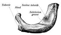
The offset rib (the topmost one) is the most curved and usually the shortest of all the ribs; it is wide and flat, its surfaces looking up and downwardly, and its borders inward and outward.
-
Get-go rib seen from in a higher place.
-

Costal groove position on a central rib.

The head is small and rounded, and possesses only a unmarried articular facet, for articulation with the body of the offset thoracic vertebra. The neck is narrow and rounded. The tubercle, thick and prominent, is placed on the outer border. It bears a small facet for articulation with the transverse costal facet on the transverse procedure of T1. There is no angle, but at the tubercle, the rib is slightly bent, with the convexity upward, so that the head of the bone is directed down. The upper surface of the body is marked past two shallow grooves, separated from each other by a slight ridge prolonged internally into a tubercle, the scalene tubercle, for the attachment of the anterior scalene; the anterior groove transmits the subclavian vein, the posterior the subclavian artery and the lowest torso of the brachial plexus. Behind the posterior groove is a crude area for the attachment of the medial scalene. The under surface is polish and without a costal groove. The outer edge is convex, thick, and rounded, and at its posterior part gives zipper to the first digitation of the serratus anterior. The inner border is concave, thin, and sharp, and marked nigh its center past the scalene tubercle. The anterior extremity is larger and thicker than that of any of the other ribs.
The 2d rib is the second uppermost rib in humans or second most frontal in animals that walk on four limbs. In humans, the second rib is divers as a true rib since information technology connects with the sternum through the intervention of the costal cartilage anteriorly (at the forepart). Posteriorly, the second rib is connected with the vertebral column by the second thoracic vertebra. The 2nd rib is much longer than the first rib, only has a very similar curvature. The not-articular portion of the tubercle is occasionally only feebly marked. The angle is slight and situated close to the tubercle. The body is non twisted so that both ends touch any plane surface upon which information technology may be laid; but there is a bend, with its convexity upwards, similar to, though smaller than that found in the first rib. The torso is non flattened horizontally like that of the first rib. Its external surface is convex, and looks up and a little outward; near the middle of information technology is a rough eminence for the origin of the lower function of the starting time and the whole of the 2d digitation of the serratus inductive; behind and to a higher place this is attached the posterior scalene. The internal surface, smooth, and concave, is directed downwardly and a little inward: on its posterior part there is a short costal groove betwixt the ridge of the internal surface of the rib and the inferior border. It protects the intercostal space containing the intercostal veins, intercostal arteries, and intercostal nerves.[half dozen] [four]
The 9th rib has a frontal part at the same level every bit the first lumbar vertebra. This level is called the transpyloric aeroplane, since the pylorus is as well at this level.[7]
The tenth rib attaches direct to the trunk of vertebra T10 instead of between vertebrae like the second through ninth ribs. Due to this directly zipper, vertebra T10 has a complete costal facet on its body.[3]
The iv floating ribs indicated
The eleventh and twelfth ribs, the floating ribs, accept a single articular facet on the head, which is of rather large size. They have no necks or tubercles, and are pointed at their anterior ends. The eleventh has a slight angle and a shallow costal groove, whereas the twelfth does not. The twelfth rib is much shorter than the eleventh rib, and its caput is inclined slightly downward.[ commendation needed ]
Sternum [edit]
The sternum is a long, flat bone that forms the forepart of the rib cage. The cartilages of the tiptop seven ribs (the true ribs) join with the sternum at the sternocostal joints. The costal cartilage of the second rib articulates with the sternum at the sternal angle making it like shooting fish in a barrel to locate.[viii]
The transversus thoracis muscle is innervated past one of the intercostal nerves and superiorly attaches at the posterior surface of the lower sternum. Its inferior attachment is the internal surface of costal cartilages two through six and works to depress the ribs.[ix]
Development [edit]
Expansion of the rib cage in males is caused by the effects of testosterone during puberty.[x] Thus, males generally have broad shoulders and expanded chests, allowing them to inhale more air to supply their muscles with oxygen.

Variation [edit]
Variations in the number of ribs occur. Nigh i in 200-500 people take an additional cervical rib, and at that place is a female predominance.[xi] Intrathoracic supernumerary ribs are extremely rare.[12] The rib remnant of the 7th cervical vertebra on i or both sides is occasionally replaced past a free actress rib called a cervical rib, which can mechanically interfere with the fretfulness (brachial plexus) going to the arm.
In several ethnic groups, most significantly the Japanese, the tenth rib is sometimes a floating rib, as information technology lacks a cartilaginous connection to the 7th rib.[3]
Function [edit]

The result of the wrinkle of the accompaniment muscles of inhalation, pulling the front of the rib cage upward, a motion known as the 'pump handle movement'. This increases the antero-posterior diameter of the thorax, contributing to the expansion in the book of the chest. A similar issue, known as the 'bucket handle movement' causes the transverse bore of the chest to increase, because non only do the ribs slant downwards from the back to the front, but, in the example of the lower ribs, also from the midline downwardly to the sides of the breast.
The human rib cage is a component of the human respiratory system. Information technology encloses the thoracic crenel, which contains the lungs. An inhalation is accomplished when the muscular diaphragm, at the floor of the thoracic cavity, contracts and flattens, while the contraction of intercostal muscles lift the rib cage upwardly and out.
Expansion of the thoracic cavity is driven in 3 planes; the vertical, the anteroposterior and the transverse. The vertical plane is extended by the assist of the diaphragm contracting and the abdominal muscles relaxing to arrange the downward pressure level that is supplied to the intestinal viscera past the diaphragm contracting. A greater extension tin exist achieved by the diaphragm itself moving down, rather than simply the domes flattening. The 2d plane is the anteroposterior and this is expanded by a movement known equally the 'pump handle.' The downward sloping nature of the upper ribs are equally such considering they enable this to occur. When the external intercostal muscles contract and lift the ribs, the upper ribs are able also to button the sternum upwardly and out. This movement increases the anteroposterior bore of the thoracic cavity, and hence aids breathing further. The tertiary, transverse, plane is primarily expanded by the lower ribs (some say it is the seventh to 10th ribs in detail), with the diaphragm's central tendon interim as a fixed betoken. When the diaphragm contracts, the ribs are able to evert (meaning turn outwards or inside out) and produce what is known as the bucket handle motility, facilitated past gliding at the costovertebral joints. In this manner, the transverse diameter is expanded and the lungs tin fill.
The circumference of the normal adult human rib cage expands by 3 to 5 cm during inhalation.[13]
Clinical significance [edit]
Rib fractures are the most common injury to the rib cage. These most frequently bear upon the middle ribs. When several next ribs incur ii or more than fractures each, this can upshot in a flail chest which is a life-threatening condition.
A dislocated rib tin can be painful and can be acquired but by coughing, or for example by trauma or lifting heavy weights.[fourteen]
One or more than costal cartilages can get inflamed – a condition known as costochondritis; the resulting pain is similar to that of a heart attack.
Abnormalities of the rib cage include pectus excavatum ("sunken chest") and pectus carinatum ("pigeon breast"). A bifid rib is a bifurcated rib, dissever towards the sternal end, and unremarkably simply affecting i of the ribs of a pair. Information technology is a congenital defect affecting about i.2% of the population. It is oft without symptoms though respiratory difficulties and other issues tin arise.
Rib removal is the surgical removal of one or more ribs for therapeutic or corrective reasons.
Rib resection is the removal of part of a rib.
Lodge and culture [edit]
The position of ribs tin be permanently altered by a form of trunk modification chosen tightlacing, which uses a corset to compress and move the ribs.
The ribs, particularly their sternal ends, are used as a way of estimating age in forensic pathology due to their progressive ossification.[15]
History [edit]
The number of ribs equally 24 (12 pairs) was noted by the Flemish anatomist Vesalius in his key work of anatomy De humani corporis fabrica in 1543, setting off a wave of controversy, as it was traditionally assumed from the Biblical story of Adam and Eve that men's ribs would number one fewer than women's.[sixteen]
Other animals [edit]

Tyrannosaurus rib cage, University of California Museum of Paleontology
In herpetology, costal grooves refer to lateral indents along the integument of salamanders. The grooves run between the axilla to the groin. Each groove overlies the myotomal septa to marking the position of the internal rib.[17] [18]
Birds and reptiles have bony uncinate processes on their ribs that project caudally from the vertical department of each rib.[19] These serve to adhere sacral muscles and also help in allowing greater inspiration. Crocodiles have cartilaginous uncinate processes.
Additional images [edit]
-
Thoracic Muzzle with Spine - Anatomy
-
Anterior surface of sternum and costal cartilages.
-

X-ray epitome of a human chest, with ribs labelled.
-

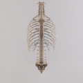
3D model of rib muzzle
-

Surface projections of the trunk, including each rib, and the costal margin.
-

Thoracic Cage with Both Humerii
See likewise [edit]
- Articulation of head of rib
- Rachitic rosary
- Terms for anatomical location
- Terms for bones
Notes [edit]
![]() This article incorporates text in the public domain from the 20th edition of Gray's Anatomy (1918)
This article incorporates text in the public domain from the 20th edition of Gray's Anatomy (1918)
- ^ "The Thoracic Cage · Anatomy and Physiology". Retrieved x March 2018.
- ^ Hyman, Libbie Henrietta (1992). Hyman's Comparative Vertebrate Beefcake. Academy of Chicago Press. p. 230. ISBN9780226870137 . Retrieved 10 March 2018.
- ^ a b c Saladin, Kenneth (2010). Anatomy and Physiology: The Unity of Form and Function. United states: The McGraw-Hill Companies, Inc. p. 485. ISBN978-0-07-352569-iii.
- ^ a b Smith, Sarah. "Intercostal spaces | Radiology Reference Commodity | Radiopaedia.org". radiopaedia.org.
- ^ http://www.teachmeanatomy.com/osteology-of-the-thorax/ [ permanent dead link ]
- ^ Moore, Dalley & Agur. 2009. Clinically Oriented Anatomy, 6th Edition. xc Pp. Lippincott, Williams & Wilkins, ISBN 0-7817-7525-6, ISBN 978-0-7817-7525-0
- ^ Bålens ytanatomi (surface anatomy). Godfried Roomans, Mats Hjortberg and Anca Dragomir. Institution for Beefcake, Uppsala. 2008.
- ^ Agur, Anne M.R.; Dalley, Arthur F. II (2009). Grant'southward Atlas of Anatomy, 12th Edition . Philadelphia, PA: Lippincott Williams and Wilkins. p. ten. ISBN978-0-7817-7055-2.
- ^ Agur, Anne M.R.; Dalley, Arthur F. 2 (2009). Grant's Atlas of Anatomy, Twelfth Edition . Philadelphia, PA: Lippincott Williams and Wilkins. p. 21. ISBN978-0-7817-7055-2.
- ^ Testosterone causes expansion of rib cage during puberty equally one of secondary sexual activity characteristics."Archived copy". Archived from the original on 2015-09-eleven. Retrieved 2013-12-31 .
{{cite web}}: CS1 maint: archived copy every bit championship (link) - ^ Kurihara Y; Yakushiji YK; Matsumoto J; Ishikawa T; Hirata 1000 (Jan–Feb 1999). "The Ribs: Anatomic and Radiologic Considerations" (PDF). RadioGraphics. Radiological Society of Due north America. 19 (1): 105–119. doi:10.1148/radiographics.19.1.g99ja02105. ISSN 1527-1323. PMID 9925395. Retrieved August 13, 2009.
- ^ Kamano H; Ishihama T; Ishihama H; Kubota Y; Tanaka T; Satoh K (June 1, 2006). "Bifid intrathoracic rib: a case study and nomenclature of intrathoracic ribs" (PDF). Internal Medicine. The Japanese Lodge of Internal Medicine. 45 (9): 627–630. doi:10.2169/internalmedicine.45.1502. PMID 16755094.
- ^ Respiratory system examination Archived 2012-03-23 at the Wayback Auto citing: Wellness & Physical Cess, Mosby-Year Book, inc. School of Nursing, Peking Academy, 2003
- ^ "Anatomy of the Human ribs - Confused Rib". Dislocated Rib. two February 2016. Archived from the original on 12 August 2016.
- ^ Franklin, D (Jan 2010). "Forensic historic period estimation in human skeletal remains: current concepts and future directions". Legal Medicine (Tokyo, Nippon). 12 (ane): 1–seven. doi:10.1016/j.legalmed.2009.09.001. PMID 19853490.
- ^ "Chapter 19 On the Bones of the Thorax". Archived from the original on 2007-07-06. Retrieved 2007-08-23 .
- ^ Duellman, W.E., Trueb, L. (1986). Biology of Amphibians. 670 Pp. McGraw - Hill Book Company, New York, New York, ISBN 0-8018-4780-Ten, 9780801847806
- ^ J. W. Petranka. 1998. Salamanders of the United States and Canada. 587 Pp. Smithsonian Institution Press, ISBN i-56098-828-2, ISBN 978-one-56098-828-1
- ^ Kardong, Kenneth V. (1995). Vertebrates: comparative anatomy, function, evolution. McGraw-Hill. pp. 55, 57. ISBN0-697-21991-7.
References [edit]
- Orientation of the intercostal muscle fibers in the human rib cage, Subit D., Glacet A., Hamzah Grand., Crandall J., Computer Methods in Biomechanics and Biomedical Engineering science, 2015, xviii, pp. 2064–2065
- Clinically Oriented Anatomy, 4th ed. Keith L. Moore and Robert F. Dalley. pp. 62–64
- Principles of Anatomy Physiology, Tortora GJ and Derrickson B. 11th ED. John Wiley and Sons, 2006. ISBN 0-471-68934-3
- De Humani Corporis Fabrica: online English translation of Vesalius' books on human beefcake.
External links [edit]
| | Wikimedia Commons has media related to Rib cage. |
Ribs And Sternum [ permanent dead link ]
- Diagram at mhhe.com
landerosbobjecied.blogspot.com
Source: https://en.wikipedia.org/wiki/Rib_cage
0 Response to "what allows the ribs to attach to the thoracic vertebrae"
Post a Comment